Perforation of Meckel Diverticulum in a Preterm Newborn
Amel Ben hamad
Published Date: 2022-04-09DOI10.36648/ippecm/22.6.027
Amel Ben hamad*
Department of Neonatology, Hedi Chaker Hospital, Sfax, Tunisia
- *Corresponding Author:
- Amel Ben hamad
Department of Neonatology, Hedi Chaker Hospital, Sfax, Tunisia
Tel: 21624658717
E-mail: benhamad.amel@gmail.com
Received Date:January 30, 2022, Manuscript No. IPPECM-22-12408; Editor assigned date: February 02, 2022, Pre QC No. IPPECM-22-12408 (PQ); Reviewed date: February 17, 2022, QC No. IPPECM-22-12408; Revised date: April 01, 2022, Manuscript No. IPPECM-22-12408 (R); Published date: April 9, 2022, DOI: 10.36648/ippecm/22.6.027
Citation: Ben hamad A (2022) Perforation of Meckel Diverticulum in a Preterm Newborn. Pediatr Emerg Care Med Open Access Vol:6 No:7
Abstract
Meckel’s diverticulum is the commonest congenital abnormality of the gastrointestinal tract. It can be complicated by hemorrhage, bowel obstruction, inflammation, perforation, intussusception, volvulus, and malignant transformation. MD perforation is rare in newborns and very rare in preterm babies. We report a case of a preterm female, born at 34 weeks gestation, with a birth weight of 1900 g admitted at birth to our intensive care unit for prematurity and hypotrophy. 48 hours after feeding, she presented rectal bleeding and green residues which necessitated diet stop. But the baby presented isolated abdominal distension after reintroducing of feeding.An abdominal radiograph showed a pneumoperitoneum. Laparotomy revealed Meckel's perforation. The baby was discharged on day 19 of life. To our knowledge, this is the only case reported with two symptoms rectal bleeding and abdominal distension.
MD is very rare but it should be considered as a differential diagnosis in every newborn presenting rectal bleeding or isolated abdominal distension.
Keywords
Meckel’s diverticulum; Perforation; Pneumoperitoneum; Prematurity
Introduction
Meckel’s diverticulum is the commonest congenital abnormality of the gastrointestinal tract [1]. It occurs approximately in 2% of the general population [2]. Complications develop in only 4% of patients with this malformation including hemorrhage, bowel obstruction, inflammation, perforation, intussusception, and malignant transformation [3].
Symptomatic MD in the neonatal period is quite rare. Perforation of Meckel diverticulum in the preterm newborn is very rare [2]. We present the case of a 34 week’s preterm female, with a spontaneous Meckel diverticulum perforation discovered on day 6 of life and we insist on considering MD as a differential diagnosis in every patient presenting rectal bleeding or isolated abdominal distension.
CaseReport
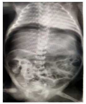
A preterm female baby was born at 34 weeks gestation by caesarian section. She was admitted to our neonatal Intensive Care Unit for low weight of birth: 1900 g. 48 hours after diet introduction, she presented rectal bleeding and green residues. Onexamination, the new born had a fever (38.5°C); her abdomen was not tender and not distended. Anal fissures diagnosis was eliminated. Enteral feeding was stopped, and we treated the patient with imipenem, amikacin, and metronidazole to cover Healthcare-associated infection. The abdominal X-ray was normal and ultrasonography of the abdomen ruled out volvulus. After 48 hours, the feeding was introduced with protein hydrolysate. The baby presented isolated abdominal distension 24 hours after feeding introduction. Abdominal X-ray showed a massive pneumoperitoneum (Figure 1).
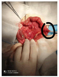
At laparotomy, a perforated Meckel's diverticulum within about 70 cm from the ileocecal valve was found. Appendicectomy was performed followed by segmental intestinal resection, removing 30 cm from the smallboweland Meckel'sdiverticulum, with end-to-end anastomosis.
On macroscopy, the affected small bowel segment was 9 × 2 cm in size and was focally dilated in which there were multiple ulcerations (Figure 2).
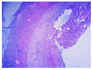
On histology, the dilated portion showed a largely ulcerated atrophic small intestine mucosa without ectopic tissue (Figure 3).
The adjacent portion of the small bowel presented a perforation with mural purulent materiel.
Our patient recovered well after surgery. She was extubated 24 hours after surgery. She received 15 days of imipenem. Enteral feeding was introduced successfully four days after surgery. The baby was discharged on day 19 of life with a regular outpatient follow-up.
Discussion
Meckel’s diverticulum is known as the most common anomaly of the digestive tract. It is a remnant of the embryonic vitelline duct that did not regress between the fifth and seventh week of gestation period when the yolk sac is replaced by the placenta [4,5].
It is considered a true diverticulum as it contains all layers of the intestine wall. In 30% to 50% of cases, heterotopic mucosa was foundwithin the diverticulum [6]. The most common ones are gastric and pancreatic tissues [7].
Ileal diverticula are one of the most common malformations of the gastrointestinal tract with an incidence ranging from 0.3% to 2.5% [8,9].
Most patients do not produce symptoms but about 2% present complications during their lives, usually at the first 2 years [10,11] such as diverticulitis [12], volvulus or obstruction [13], bleeding [14], and perforation [15].
Symptomatic DM is rare in newborns. It represents less than 20% of all children's cases. Bowel obstruction is the most common symptom at this age. The Meckel's diverticulum is revealed by a perforation in 10% of cases during the first year of life but it is exceptional in the neonatal period with only 14 cases reported during the last 30 years including 8 cases in preterm babies [16,17]. Thus, this case is the 15th reported in the literature in newborns and the 9th in premature infants. Rectal bleeding has been reported in one case [2].
The last 25 years overview of MD perforation showed that up to 75% of cases are due to inflammation or ectopic mucosa within the diverticulum [16]. Only inflammation was reported in our case, there was no heterotopic tissue.
It should be considered that intestinal perforation in neonates has other causes which include necrotizing enter colitis, intestinal obstruction secondary to Hirschsprung’s disease, intestinal atresia, or intestinal volvulus [16]. To our knowledge, this is the only patient who presents two symptoms: early bleeding and after that abdominal distension.
Several factors are recognized as predisposing for perforation in the newborn such as hypoxia, antenatal and postnatal corticosteroids therapy, exchange transfusion, and poor intrauterine blood ?ow [16,18]. Our patient was symptomatic at birth and had not any factor.
There are only two reports of perforated Meckel’s diverticulum mortality. The first occurred in 1927 when a 4-day old newborn with abdominal distension and shock died and on autopsy assessment, a perforated MD was diagnosed. The second case was a newborn baby with multiple birth defects (esophageal atresia, tracheoesophageal fistula, and imperforation of the anus) and he died of cardiorespiratory failure [1]. Our patient was discharged after 19 days.
Conclusion
Although MD perforation is rare in the neonate period, it should be remembered as a cause of acute abdomen. Early recognition and prompt surgery are necessary for a positive outcome. The early two symptoms of our patient are the originality of this reported case.
References
- Frooghi M, Bahador A, Golchini A, Haghighat M, Ataollahi M, et al. (2016) Perforated Meckel’s Diverticulum in a 3-day-old Neonate; A Case Report. J Dig DisOct 8:323-326
- Borgi A, Bouziri A, Boujelbene N, Sghairoun N, Belhadj S, et al. (2014) Perforated Meckel’s Diverticulum in a Very Preterm Baby Revealed at Birth. Fetal Pediatr Pathol 33:119-122
- Perforated Meckel’s Diverticulum in a 3-day-old Neonate; A Case Report
- Agha RA, Franchi T, Sohrabi C, Mathew G (2020) The SCARE 2020 Guideline: Updating Consensus Surgical CAseREport (SCARE) Guidelines. Int J Surg 84 226–230.
- Soltero MJ, Bill AH (1976) The natural history of Meckel’s diverticulum and its relation to incidental removal. Am J Surg 132:168–171
- Elsayes KM, Menias CO, Harvin HJ (2007) Imaging manifestations of Meckel’s diverticulum. AJR Am J Roentgenol 189:81–88
- Martin JP, Connor PD, Charles K (2000) Meckel’s diverticulum. Am Fam Physcian 15 :1037–1042
- Sameer Mohiuddin S, Gonzalez A, Corpron C (2011) Meckel’s Diverticulum with Small Bowel Obstruction Presenting as Appendicitis in a Pediatric Patient. JSLS 15:558-561
- Francis A, Kantarovich D, Khoshnam N, Alazraki AL, Patel B, et al. (2016) Pediatric Meckel's Diverticulum: Report of 208 Cases and Review of the Literature. Fetal and Pediatric Pathology 35:199-206
- Huang CC, Lai MW, Hwang FM, Yeh YC, Chen SY, et al. (2014) Diverse presentations in pediatric Meckel’s diverticulum: a review of 100 cases. Pediatr Neonatol 55:369–375
- Sta?nescu GL, Ple¸sea IE, Diaconu R, Gheonea C, Sabetay C, et al. (2014) Meckel’s diverticulum in children, clinical and pathological aspects. Rom J Morphol Embryol 55:1167–1170
- Sarli L, Costi R (2001) Laparoscopic resection of Meckel’s diverticulum: report of two cases. Surg Today 31:823–825
- Catarci M, Zaraca F, Scaccia M, Gossetti F, Negro P, et al. (1995) Laparoscopic management of volvulated Meckel’s diverticulum. Surg Laparosc Endosc 5:72–74
- Imdahl A (2001) Genesis and pathophysiology of lower gastrointestinal bleeding. Langenbecks Arch Surg 386:1–7
- Schmid SW, Schafer M, Krahenbuhl L, Buchler MW (1999) The role of laparoscopy in symptomatic Meckel’s diverticulum. Surg Endosc 13:1047–1049
- Aguayo P, Fraser JD, St Peter SD, Ostlie DJ (2009) Perforated Meckel’s diverticulum in a micropremature infant and review of the literature. Pediatr Surg Int 25:539–541.
- Crossref
- Bindi E, Cruccetti A, Ilari M, Mariscoli F, Carnielli VP, et al. (2020) Meckel's diverticulum perforation in a newborn positive to Sars-Cov – 2. J Pediatr Surg Case Rep 62:101641
Open Access Journals
- Aquaculture & Veterinary Science
- Chemistry & Chemical Sciences
- Clinical Sciences
- Engineering
- General Science
- Genetics & Molecular Biology
- Health Care & Nursing
- Immunology & Microbiology
- Materials Science
- Mathematics & Physics
- Medical Sciences
- Neurology & Psychiatry
- Oncology & Cancer Science
- Pharmaceutical Sciences
