Pediatric Detection of Cutaneous Larva Migrans on Point of Care Ultrasound
Dr. Daniella Lamour*
Published Date: 2025-02-19Daniella Lamour*
Department of Pediatrics, Harvard University, Cambridge, United States
- *Corresponding Author:
- Daniella Lamour
Department of Pediatrics, Harvard University, Cambridge, United States
E-mail:daniellalamour@gmail.com
Received date: December 09, 2023, Manuscript No. IPPECM-23-18191; Editor assigned date: December 11, 2023, PreQC No. IPPECM-23-18191 (PQ); Reviewed date: December 25, 2023, QC No. IPPECM-23-18191; Revised date: February 12, 2025, Manuscript No. IPPECM-23-18191 (R); Published date: February 19, 2025, DOI: 10.36648/IPPECM.10.1.002
Citation: Lamour D (2025) Pediatric Detection of Cutaneous Larva Migrans on Point of Care Ultrasound. Pediatr Emerg Care Med Open Access Vol:10 No:1
Abstract
Larva migrans is a cutaneous parasitic infection that occurs when an immature hookworm larva inadvertently penetrates the dermis of a human, typically on the extremities. Traditionally, this diagnosis is made clinically when a tortuous/serpiginous eruption is seen superficially in the skin with complaints of intense pruritus. Point of Care Ultrasound (POCUS) is a diagnostic tool useful in soft tissue complaints in the Emergency Department (ED). We present a case of an 18 years old female who presented to the ED with foot pruritis four days after walking on the beach barefoot. POCUS revealed several motile structures in the dermis of the patient’s foot, further confirming our suspicion of cutaneous larva migrans. The patient was then placed on an oral anthelmintic and her symptoms resolved shortly after.
Keywords
Cutaneous larva migrans; Point of care ultrasound; Soft tissue ultrasound
Introduction
Cutaneous Larva Migrans (CLM) is a common zoonotic infection that occurs when the filariform larva of the hookworm penetrates the epidermis of a human’s skin. The adult hookworm usually lays its eggs in their natural hosts, cats and dogs [1]. The most common species of these hookworms are Ancylostoma brasiliense (cat hookworm) and Ancylostoma caninum (dog hookworm) and are commonly seen in the Southern United States, the Caribbean, and South America. The prevalence has been reported up to 8% in certain populations, most commonly present in children [2]. Risk factors include a young age (10-14 years old), male sex, resource-poor region, and walking barefoot [2]. These organisms thrive in warm and moist environments, conventionally present amongst travelers in tropical regions.
Humans are inadvertent hosts, typically when walking barefoot and accidentally stepping on the contaminated animal feces infested with the hookworm. The hookworm is unable to travel into deeper layers of the skin to complete its life cycle in a human’s gastrointestinal tract, due to a deficiency in the collagenase enzyme [3]. Thus they migrate throughout the epidermis, creating the classic superficial serpiginous tracks, which may last a few weeks to months, and oftentimes fully resolve without treatment [4]. Without an objective diagnostic tool, this classically remains a clinical diagnosis. POCUS is a well established tool which has been shown to increase diagnostic accuracy in soft tissue infections in pediatric emergency medicine [5]. The most common uses of soft tissue ultrasound include cellulitis, abscesses, and foreign bodies. When there is an increased suspicion of a foreign body, Ultrasound (US) can be used to detect, localize, and potentially extract it [6]. CLM, a common parasitic infection which may be considered a foreign body, has scarcely been reported on POCUS. We present a case of CLM confirmed on POCUS in a tertiary pediatric emergency department.
Case Presentation
An 18 years old female with a history of diabetes mellitus type 1 presented with pain, itching, and swelling of the left heel for one day. Her symptoms worsened with weight-bearing and improved with rest. She stated she was at the beach four days prior but denied any injury or having stepped on anything sharp. Her vital signs were normal. The physical examination was normal with the exception that the soft tissue exam revealed an erythematous raised serpiginous eruption on the heel that was mildly tender to palpation without fluctuance, induration, or swelling. POCUS was performed to further examine the serpiginous lesion and to rule out the presence of a foreign body or abscess. The US scan revealed a serpiginous motile structure in the dermal layer of the foot, further confirming the diagnosis of CLM. The patient was given a prescription for a one-time dose of Ivermectin with improvement of symptoms.
Ultrasound findings
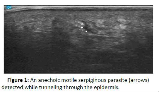
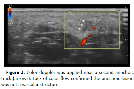
A high-frequency linear probe on a Sonosite PX US machine was placed on the plantar aspect of the patient’s foot to obtain the superficial images in B-mode. The images revealed several small linear hyperechoic lesions in the subcutaneous layers of the foot exhibiting serpiginous motility (Figure 1). The subcutaneous tissue shifted throughout the migration process. Color doppler was applied to confirm the moving echogenic lesions were not vasculature structures (Figure 2). The contralateral foot was also scanned for comparison and there was an absence of these motile structures in the epidermis.
Figure 1: An anechoic motile serpiginous parasite (arrows) detected while tunneling through the epidermis.
Figure 2: Color doppler was applied near a second anechoic track (arrows). Lack of color flow confirmed the anechoic lesion was not a vascular structure.
Discussion
Cutaneous larva migrans can be diagnosed solely on a patient’s clinical presentation, however, additional studies may help confirm the diagnosis. Serological tests, such as enzyme linked immunosorbent assay, can detect specific antibodies against the causative parasites [7]. Skin biopsy can be performed as well to identify the presence of larvae in the skin, but it may be inconclusive, rather invasive, and unnecessary. In other cases, imaging studies like X-rays may be employed to visualize the larvae in visceral tissues [8].8 Optical coherence tomography has been reported to be an effective minimally invasive tool to rapidly detect CLM [9]. However, it isn’t widely available in the ED and is rarely used outside of an ophthalmology office. Although dermoscopy has been used in the detection of these cutaneous parasites, most EDs do not carry these devices. In a recent study, reflectance confocal microscopy confirmed a larva burrow, described as a hyporeflective disruption of the normal honeycomb pattern in the epidermis [6]. As one can imagine, these devices are more commonly found in dermatology offices and are not typically stocked in most EDs. Interestingly, a high frequency US was also utilized in that same study. A cylindrical mass and shadowing were revealed which the authors believed may have corresponded to the parasite and larva burrow [6].
There are limited studies demonstrating cutaneous larva migrans with minimally invasive imaging tools in pediatric patients in the ED. In contrast to other imaging modalities,POCUS is a valuable noninvasive portable imaging tool, available in most EDs. It has many clinical applications, including distinguishing between different soft tissue complaints. This case report features a POCUS detection of CL. We highlighted motile hyperechoic lesions in the epidermal layer of the patient’s foot, utilizing a high-frequency linear transducer. When these hookworms tunnel through the skin, their paths are highlighted as anechoic tracks amid the echogenic base representing the dermis. Unfortunately, the individual larvae were not detected. While visualizing the actual larva may require more effort and time, the parasitic tracks can be detected via US, as seen with our patient. This technique may prove to be even more clinically useful as another case demonstrated that suspected larva diagnosed via US imaging, were actually normal soft tissue [6]. As more clinician’s implement POCUS when suspicion for CLM exists, there will be more images available to compare our images against and perhaps give insight to diagnostic criteria for CLM.
Our patient complained of swelling and pruritus on the sole of her foot upon her presentation to the emergency department. The typical presentation of CLM consists of tortuous, erythematous lesions which are very pruritic. However, patient presentation and symptoms are variable and CLM may be easily misdiagnosed due to lack of recognition and/or confidence in the diagnosis. As presented in our case, POCUS may serve as a useful confirmatory tool prior to initiating treatment of CLM with oral anthelmintics. The first line recommendation for anthelmintics is a one-time dose 200 mcg/kg oral Ivermectin, which usually results in a 100% cure rate. Albendazole may be used second line if necessary. Topical anthelmintics like Thiabendazole may also be effective if infection is local but has been reported to have a poor eradication rate. Our patient received one dose of Ivermectin with full resolution of her symptoms. As CLM is very responsive to treatment, early and accurate diagnosis via US evaluation is likely clinically valuable [10].
Conclusion
The diagnosis of CLM historically has been made by history and physical. Other suggested imaging modalities are either cost restrictive, insufficient, or not readily available. In this case, POCUS helped confirm the diagnosis and is not hindered by the same limitations of other imaging modalities when assessing for CLM. Further studies are needed to confirm the utility of POCUS to aid in the diagnosis of parasitic infections.
References
- Lamour D, Farrow RA, Pierre J, Khalil P (2024) Diagnosis of cutaneous larva migrans using point of care ultrasound. POCUS J 9:33-35
[Crossref] [Google Scholar] [PubMed]
- Reichert F, Pilger D, Schuster A, Lesshafft H, Guedes de Oliveira S, et al. (2018) Epidemiology and morbidity of Hookworm-related Cutaneous Larva Migrans (HrCLM): Results of a cohort study over a period of six months in a resource-poor community in Manaus, Brazil. PLoS Negl Trop Dis 12:e0006662
[Crossref] [Google Scholar] [PubMed]
- Edelglass JW, Douglass MC, Stiefler R, Tessler M (1982) Cutaneous larva migrans in northern climates: A souvenir of your dream vacation. J Am Acad Dermatol 7:353-358
[Crossref] [Google Scholar] [PubMed]
- Campoli M, Cortonesi G, Tognetti L, Rubegni P, Cinotti E (2022) Noninvasive imaging techniques for the diagnosis of cutaneous larva migrans. Skin Res Technol 28:374
[Crossref] [Google Scholar] [PubMed]
- Chen KC, Lin AC, Chong CF, Wang TL (2016) An overview of point-of-care ultrasound for soft tissue and musculoskeletal applications in the emergency department. J Intensive Care 4:1-11
[Crossref] [Google Scholar] [PubMed]
- Rooks VJ, Shiels III WE, Murakami JW (2020) Soft tissue foreign bodies: A training manual for sonographic diagnosis and guided removal. Clin Ultrasound 48:330-336
[Crossref] [Google Scholar] [PubMed]
- Kwon IH, Kim HS, Lee JH, Choi MH, Chai JY, et al. (2003) A serologically diagnosed human case of cutaneous larva migrans caused by Ancylostoma caninum. Korean J Parasitol 41:233
[Crossref] [Google Scholar] [PubMed]
- Sakai S, Shida Y, Takahashi N, Yabuuchi H, Soeda H, et al. (2006) Pulmonary lesions associated with visceral larva migrans due to Ascaris suum or Toxocara canis: Imaging of six cases. Am J Roentgenol 186:1697-1702
[Crossref] [Google Scholar] [PubMed]
- Morsy H, Mogensen M, Thomsen J, Thrane L, Andersen PE, et al. (2007) Imaging of cutaneous larva migrans by optical coherence tomography. J Travel Med. 5:243-246
[Crossref] [Google Scholar] [PubMed]
- Ogueta I, Navajas-Galimany L, Concha-Rogazy M, Alvarez-Veliz S, Vera-Kellet C, et al. (2019) Very high and high frequency ultrasound features of cutaneous larva migrans. J Med Ultrasound 38:3349-3358
[Crossref] [Google Scholar] [PubMed]

Open Access Journals
- Aquaculture & Veterinary Science
- Chemistry & Chemical Sciences
- Clinical Sciences
- Engineering
- General Science
- Genetics & Molecular Biology
- Health Care & Nursing
- Immunology & Microbiology
- Materials Science
- Mathematics & Physics
- Medical Sciences
- Neurology & Psychiatry
- Oncology & Cancer Science
- Pharmaceutical Sciences


