Breakage of Growth Hormone Needle in Subcutaneous Tissue of a Six-Year-Old Child's Buttock
Shuichi Shimada, Asako Ichihara, Masatoshi Jinnin, Satoshi Fukushima, Hiroyo Mabe,Koichi Kawanaka and Hironobu Ihn
1Department of Dermatology and Plastic Surgery, Faculty of Life Sciences, Kumamoto University, 1-1-1 Honjo, Kumamoto, 860-8556, Japan
2Kumamoto University Hospital Department of Pediatrics, Kumamoto University, 1-1-1 Honjo, Kumamoto, 860-8556, Japan
- *Corresponding Author:
- Asako Ichihara
Department of Dermatology and Plastic Surgery
Faculty of Life Sciences, Kumamoto University
1-1-1 Honjo, Kumamoto, 860-8556
Japan
Tel: 81-96-373-5233
E-mail: shimashu_p@yahoo.co.jp
Received date: October 06, 2017; Accepted date: October 10, 2017; Published date: October 14, 2017
Citation: Shimada S, Ichihara S, Jinnin M, Fukushima S, Mabe H, et al. Breakage of Growth Harmone Needle in Subcutaneous Tissue of a Sixyear- Old Child's Buttock. Pediatr Emerg Care Med Open Access. 2017, Vol.2 No.1:2.
Copyright: © 2017 Shimada S, et al. This is an open-access article distributed under terms of the Creative Commons Attribution License, which permits unrestricted use, distribution, and reproduction in any medium, provided the original author and source are credited.
Abstract
Here, we present a six-year-old boy, whose growth hormone (GH) injection needle (34-gauge, 4 mm length) broke and was embedded in the subcutaneous tissue of his buttock. First, we immediately attempted to remove the needle under local anesthesia, however, we were unable to locate the needle due to its tiny size even though we used ultrasonography. Therefore, we used computed tomography (CT) guided marking to remove the needle under general anesthesia. We want to emphasize that patients receiving GH therapy should be aware of the possibility of needle breakage, and they should be taught proper injection techniques to prevent accidents.
Keywords
Growth hormone; Needle-breakage; Computed tomography (CT) guided marking; Injection needle
Introduction
Growth hormone (GH) has been used for the treatment of growth disorders such as hypopituitarism and hereditary disorders for more than 50 years [1]. The standard method for growth hormone (GH) administration is subcutaneous injection daily into the patient’s buttock or thigh [1]. To reduce pain at the injection site, shorter and thinner needles have been developed. Currently in Japan, injection pens with a thin (usually 32-gauge to 34-gauge) disposal needles are often used, especially for younger patients. The injections are usually performed by the parents after they are educated on the appropriate injection techniques.
There have been several reports of injection needle breakage duringa insulin dministration by patients themselves or during dental or local anesthesia administration [2,3]. However, only one case of needle breakage during a GH injection has been reported [4]. We present a six-year-old boy whose GH injection needle broke and embedded in the subcutaneous tissue of his buttock, resulting in the successful removal operation using computed tomography (CT) guided marking.
Case Report
A six-year-old boy, suspected to have a broken GH needle (34-gauge, length 4 mm) embedded in his left buttock, presented to our clinic on the day following GH administration. The patient had a history of hypopituitarism and had been receiving GH injection therapy for a few years. The injection was performed by his mother for years without any incident so far. However, according to his mother, the needle broke at the root when the patient suddenly moved during the injection. She had injected GH while he was sleeping, which caused him to move suddenly in surprise.
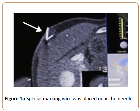
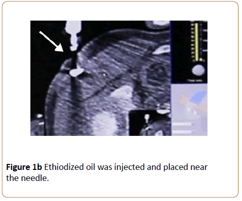
Physical examination showed a needle mark with erythema on his left buttock. His laboratory findings were normal. Radiograph and ultrasound images of his buttock revealed the presence of the broken tiny needle in the subcutaneous tissue, though the images were not clear enough to detect the location. We attempted surgical removal under local anesthesia, however, we were unable to find the small needle. Next, we performed an operation under general anesthesia. Though we intended to perform the operation immediately, we operated one week later after all. The location of the embedded needle was not clear enough in the preoperative xray image. Therefore, preoperative CT-guided marking was performed by an interventional radiologist. As the CT image showed the embedded needle under the skin, a special marking wire for lung tumors was placed near the needle. Ethiodized oil was injected near the needle so that the special marking wire and the ethiodized oil mass can serve as markers for the embedded needle (Figure 1a and 1b).
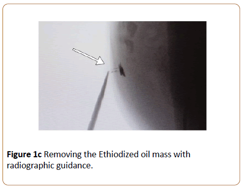
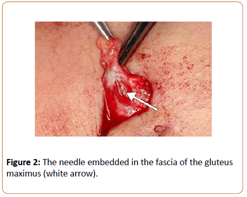
Right after the CT-guided marking, we started the operation under general anesthesia. We could find the ethiodized oil mass easily inside the subcutaneous fat and removed it using radiographic guidance (Figure 1c). We found the needle was embedded in the fascia of the gluteus maximus, just below the ethiodized oil mass (Figure 2). The broken needle was excised and the postoperative course was uneventful.
Discussion
GH needle breakage is a rare complication, with only one case reported so far [4]. As the number of patients with diabetes increases, more patients are using injection pens. GH needles are similar to ones used for insulin administration, with similar risk of adverse events.
However, the number of potential accidents is estimated to be higher than that of incidents actually reported. It is likely that the number of adverse events may continue to increase in the future. Medical accidents can be caused by two primary factors; medical devices and patients. Injection needles used in Japan are fine and thin (32-gauge to 34- gauge) to minimize pain. In this case, the thinnest 34-gauge needle (0.18 mm in diameter) was used. Since these injection needles are made very fine, they are easily broken by lateral external forces [5]. Moreover, since the needle itself is difficult to see, there are cases in which patients accidentally puncture their fingers or use the bending needles. Incompetent injections including the wrong insertion angle or changing the syringe angle during the injection can lead to the needle breakage. Furthermore, avoiding sudden or unexpected movement is required, especially in children. In this case, the patient’s sudden movement was the primary cause of the needle breakage.
After this incident, the manufacturer performed a product testing again. According to the test report, when lateral external force was applied to the needle, it broke in the same way as this case.
When subcutaneous foreign matter is non-permeable to radiographs as in this case, it is generally easy to identify and remove [6]. However, when tiny objects such as injection needles are embedded deeply in subcutaneous tissue, such as the buttocks, it is very difficult to identify and remove them even under x-ray or ultrasonography.
In this case, the needle was embedded in the buttock, and it was difficult to grasp the three-dimensional location, even under ultrasonography or radiograph. In addition, since this was a pediatric case, we decided to perform CT guided marking to remove urgently and minimize risk of general anesthesia due to hypopituitarism. The interventional radiologist used marking wire, which has a small hook on the end and is usually used for preoperative marking of lung tumor surgery. They also use ethiodized oil for preoperative marking so that the ethiodized oil mass would be a marker during the operation [7].
In this case, it was a very thin and short needle (length of 4 mm). Fortunately, the marking wire and ethiodized oil were successfully placed close to the injection needle. Removal of the embedded needle became easier with the preoperative marking.
There have been no reports that discuss the removal of a subcutaneous foreign body under CT-guided marking. If the needle migrates into the blood vessels, severe complications can ensue. The needle has the potential to embolize vessels of the heart or lungs [8]. Prompt removal is therefore necessary [9]. As ultrasonography techniques have developed, there have been many reports on detecting and removing subcutaneous foreign bodies under ultrasonography [10]. Ultrasonography is becoming the most useful and harmless method [11,12]. In this case, at first we performed operation under local anesthesia with ultrasonography. However, it was very difficult to detect a very thin needle near the muscle in a short period of time during the child can be patient. In cases where the foreign body is very small and located in the parts like a buttock, preoperative ethiodized oil marking under CT is a useful and safe procedure [7,13]. Since the detection and removal of a soft-tissue foreign body (especially a thin needle) are often very difficult, preoperative marking is recommended. Patients receiving GH therapy should be aware of the possibility of needle breakage and should learn proper techniques to prevent adverse events.
References
- Robert K, Anna GD, Anna BC, Bogusaw O (2007) Growth hormone therapy in children and adults. Pharmacol Rep 59: 500-516.
- Augello M, von Jackowski J, Gratz KW, Jacobsen C (2011) Needle breakage during local anesthesia in the oral cavity-a retrospective of the last 50 years with guidelines for treatment and prevention. Clin Oral Investig 15: 3-8.
- Sood A, Miglani S, Moorthy D (2001) Breakage of Insulin Syringe Needle in Subcutaneous Tissue. J Pediatr Endocrinol Metab 14: 101-102.
- Kim SH, Huh K, Jee YS, Park MJ (2011) Breakage of growth hormone needle in subcutaneous tissue. J Spec Pediatr Nurs 16: 162-165.
- American Diabetes Association (2003) Insulin administration. Diabetes Care 26: 121-124.
- Ginsburg MJ, Ellis GL, Flom LL (1990) Detection of soft-tissue foreign bodies by plain radiography, xerography, computed tomography, and ultrasonography. Ann Emerg Med 19: 701-703.
- Kawanaka K, Nomori H, Mori T, Ikeda K, Ikeda O, et al. (2009) Marking of small pulmonary nodules before thoracoscopic resection: Injection of Lipiodol under CT-fluoroscopic guidance. Acad Radiol 16: 39-45.
- Woodhouse JB, Uberoi R (2012) Techniques for intravascular foreign body retrieval. Cardiovasc Intervent Radiol 36: 888-897.
- Ngaage DL, Cowen ME (2001) Right ventricular needle embolus in an injecting drug user: the need for early removal. Emerg Med J 18: 500-501.
- Borgohain B, Borgohain N, Handique A, Gogoi PJ (2012) Case report and brief review of literature on sonographic detection of accidentally implanted wooden foreign body causing persistent sinus. Crit Ultrasound J 4: 10-11.
- Oikarinen KS, Nieminen TM, Makarainen H, Pyhtinen J (1993) Visibility of foreign bodies in soft tissue in plain radiographs, computed tomography, magnetic resonance imaging, and ultrasound: An in vitro study.Int J Oral Maxillofac Surg 22 :119-124.
- Aras MH, Miloglu O, Barutcugil C, Kantarci M, Ozcan E, et al. (2010) Comparison of the sensitivity for detecting foreign bodies among conventional plain radiography, computed tomography and ultrasonography. Dentomaxillofacial Radiol 39: 72-78.
- Katada K, Kato R, Anno H, Ogura Y, Koga S, et al. (1996) Guidance with real-time CT fluoroscopy: early clinical experience. Radiology 200: 851-856.

Open Access Journals
- Aquaculture & Veterinary Science
- Chemistry & Chemical Sciences
- Clinical Sciences
- Engineering
- General Science
- Genetics & Molecular Biology
- Health Care & Nursing
- Immunology & Microbiology
- Materials Science
- Mathematics & Physics
- Medical Sciences
- Neurology & Psychiatry
- Oncology & Cancer Science
- Pharmaceutical Sciences
